

Approved products, models and indications may differ from country to country.
Please contact us for detail.
| Generic name | β-tricalcium phosphate |
|---|---|
| Composition | Ca3(PO4)2 |
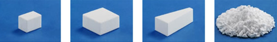
| Appearance | White porous material |
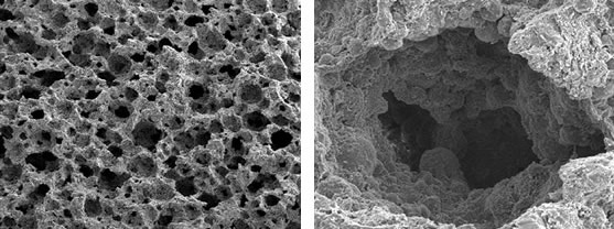
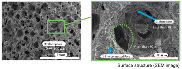
SUPERPORE has triple pore structure that is suitable for bone regeneration as illustrated in APACERAM Type-AX.
Evaluation by short to long term implantation test in canines.※3
As a result, regeneration of the bone was achieved in both cortical bone and medullary cavity including anatomical shape.
SUPERPORE high porosity type blocks (porosity 75%, Ø4mm x 12mmL) were implanted in the femurs of the canines and extracted in 4, 8, 13 and 26 weeks. Samples stained by toluidine blue were prepared for observation. (Implantation test was executed in accordance to ISO10993 in the facility of GLP level.)
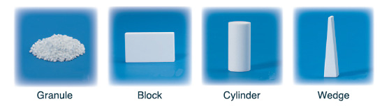
Granules, blocks, and cylinder models are available.
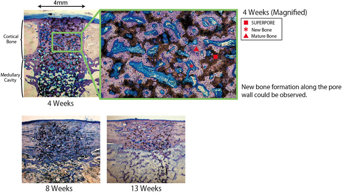
SUPERPORE balanced type blocks (porosity 67%, Ø4mm x 12mmL) were implanted in the femurs of canines and extracted in 4 and 13 weeks. Samples stained by toluidine blue were prepared for observation. Implantation tests were executed in accordance to ISO10993 in the facility of GLP level.
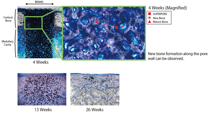
Active bone formation was observed around the material in 4 weeks. Early stage of the bone formation was observed in the pores whileabsorption of the SUPERPORE (Balanced type) l and replacement of new bone tissues were observed immediately below the periosteum and at the interface of the medullary cavity.
In 13 weeks, bone formation continuing the host bone and absorption of the material were observed in the cortical area, while medullary cavity and adipose tissues were formed in the medullary cavity area.
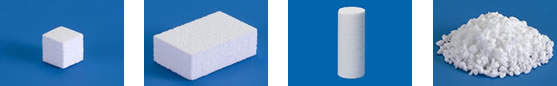
Granules, blocks, cylinder and wedge models are available.
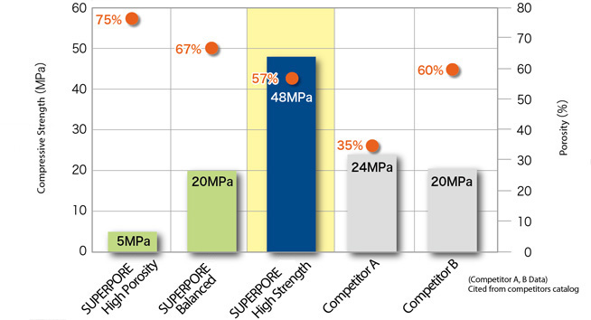
3. High Strength type(porosity 57%)
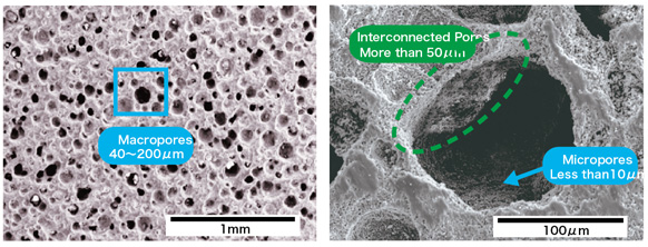
| 【Structure】 | 【Form】 | |
| Triple pore structure | Macro pores | 40〜200μm |
| Interconnecting pores | ≥40μm | |
| Micro pores | ≤10μm |
SUPERPORE high strength type blocks (porosity 57%, Ø4mm x 12mmL) were implanted in the femurs of the beagles and extracted in 26 and 52 weeks. Undecalcified samples stained by Hematoxylin and Eosin were prepared for observation. Implantation test was executed in accordance to ISO10993 in the facility of GLP level.
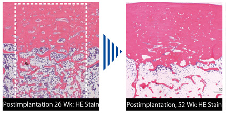
26 weeks after implantation:Bone formation continuing to host bone and absorption of the material and replacement by bone were observed in the cortical area. 52 weeks after implantation: The material was almost absorbed and replaced by bone. Bone regeneration was observed allover the implantation area including anatomical form.
Granules, blocks, cylinder and wedge models are available.